


Seeds

Last week’s announcement of my role as the UK’s first Blue Carbon Artist in Residence was followed by a flutter of meetings, conversations, and emails. Friday found me walking around Plymouth from meeting to meeting, from the Aquarium to the University to the Marine Station. Each meeting was for a different part of the project, a different piece of the idea. I caught up with my project partners, picked up seed samples and dropped them off, and had a hugely encouraging conversation about filming, photography, and boat trips.
Every meeting took me one step further, moving me from the having ideas stage to the making these ideas a reality stage of this residency. That reality truly began today with the creation of the first images for the project.
These images where of seagrass seed samples generously given to me by ReMEDIES at the Ocean Conservation Trust, who are working on seagrass restoration in Plymouth Sound. On Friday afternoon I dropped them off at Plymouth Universities Electron Microscopy Centre to be prepared.
I returned this morning to study and photograph the samples through the Scanning Electron Microscope. Or more accurately to sit and help with this, as it was the wonderful technician Glenn Harper who worked the microscope. After all, I would have no idea what to do.
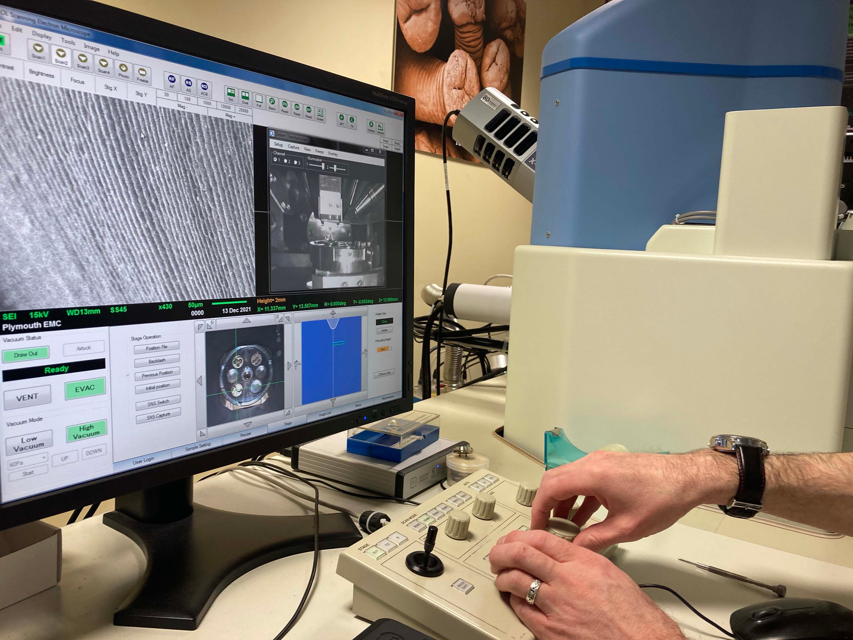
The microscope can zoom in so close everything we see is measured in micrometres, which are 1000th of a millimetre. The specimens need to conduct electricity and so my seeds were coated in gold. The electron microscope forms the image by scanning secondary electrons. The specimens sit in a vacuum chamber. If you don’t know what half of that means don’t worry, neither do I.
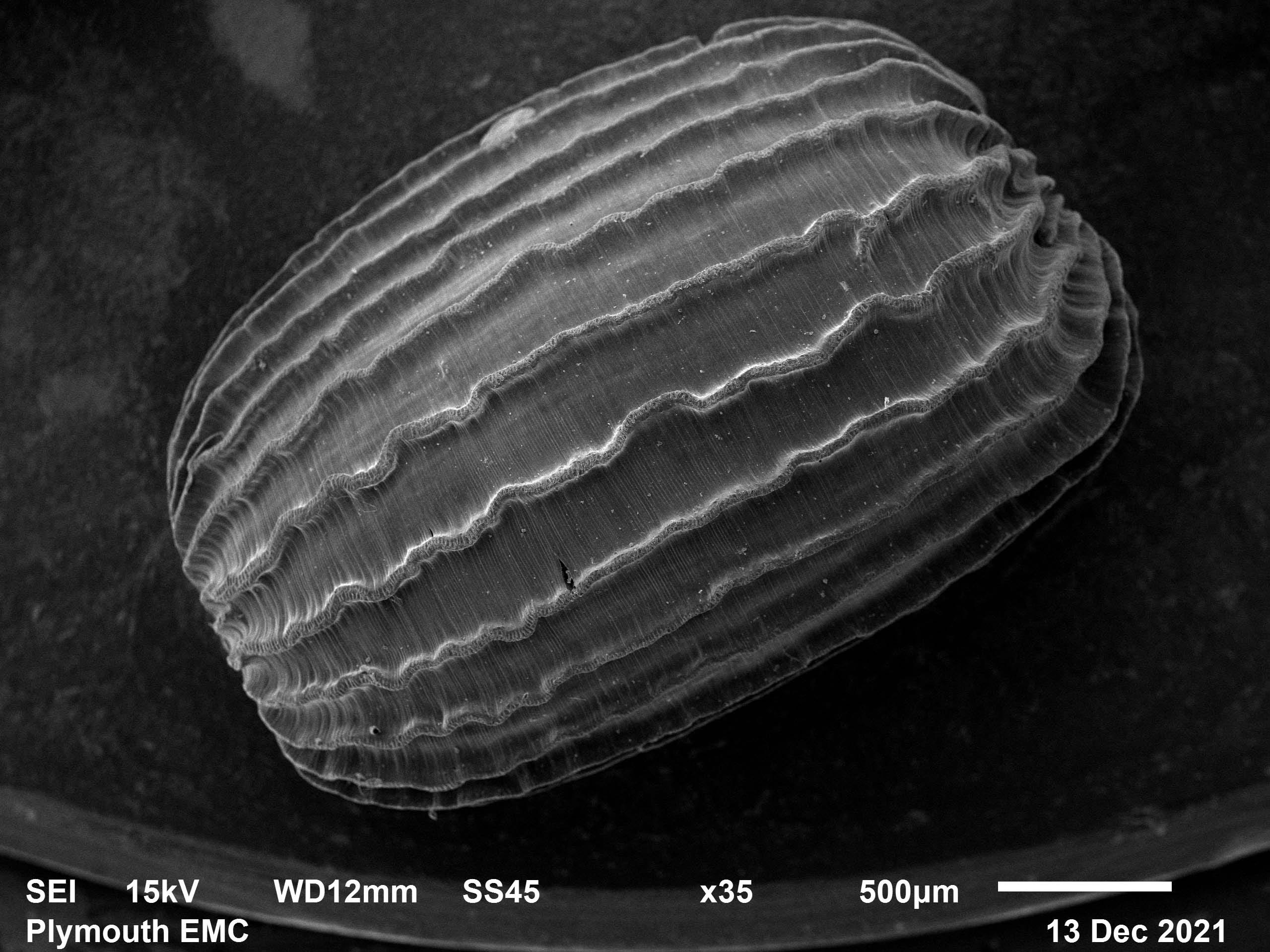
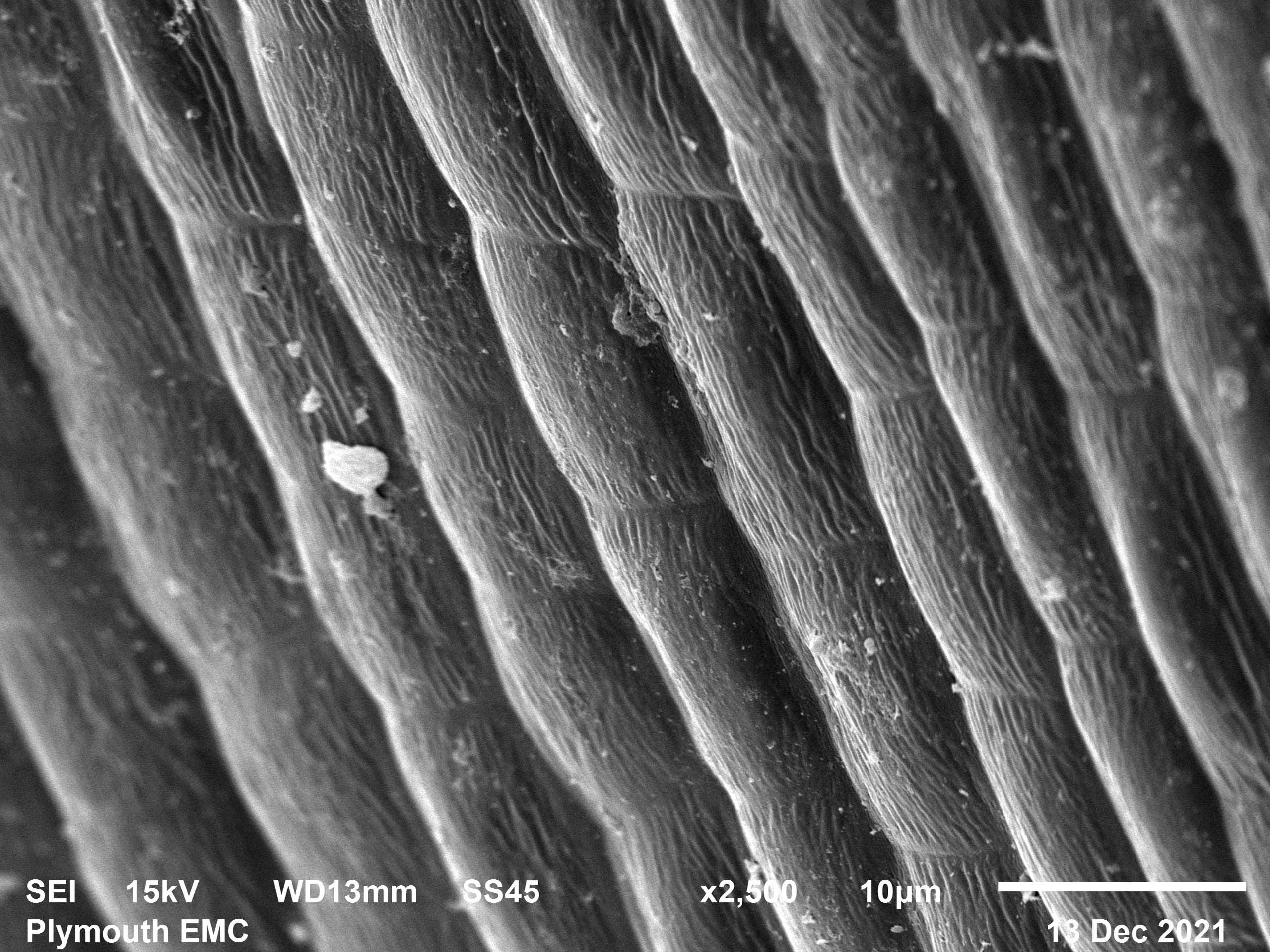
What I do know is that today I saw and photographed the individual cells of a single seagrass shoot, and four separate seeds. They were truly stunning.
I have a lot of ideas, all of which are still too fuzzy to share yet, but I promised you lovely subscribers a behind the scenes look so I shall see if I can make sense of one of them for you.
The space I am taking over in the aquarium is a long corridor. My plan is to break the space up with sculptural structures that make visitors weave through the space rather than walk, or in the case of most of the kids, run, directly down it without stopping.

Originally I had imagined something seagrass shaped, and then I attended an online event on electron microscopy and had a better idea. My hope with visiting the PEMC today was that the images seen through the microscope might form the foundation of these structures. So far, I think I’m in luck. I am going back with more samples in the new year, but even these first images suggest that this incredible plant’s cellular structure could easily be interpreted into something sculptural.
When I began this project, I decided I wanted to use as many scientific techniques as possible to make the eventual work. This is the first technique used and it’s exciting to know so early on that my planned approach can result in such incredible and rich imagery. I look forward to sharing more with you and discovering together if my ideas come together.
For anyone who wants to know more about Electron Microscopy the team at Plymouth Uni do free live events which are fascinating- and the next one is Christmas themed! Book HERE
Enjoying A Nomadic Rose, why not share it?